
Built by a practicing radiologist
Ultrasound reports, at the speed of your voice.
Dictate your findings and get a polished, branded report — structured by the platform's standard exam models, with calculators, classifications and reference tables in the same window.
No install · works in your browser · full access during the trial
Voice, AI-text or manual
Calculators in the report
Branded PDF + QR seal
10 languages
sonoaireport.com — reporting
9.4hsaved per week
160+calculators & references
10languages
1window, end to end
The dictation engine
The best ultrasound dictation on the market — engineered by a specialist.
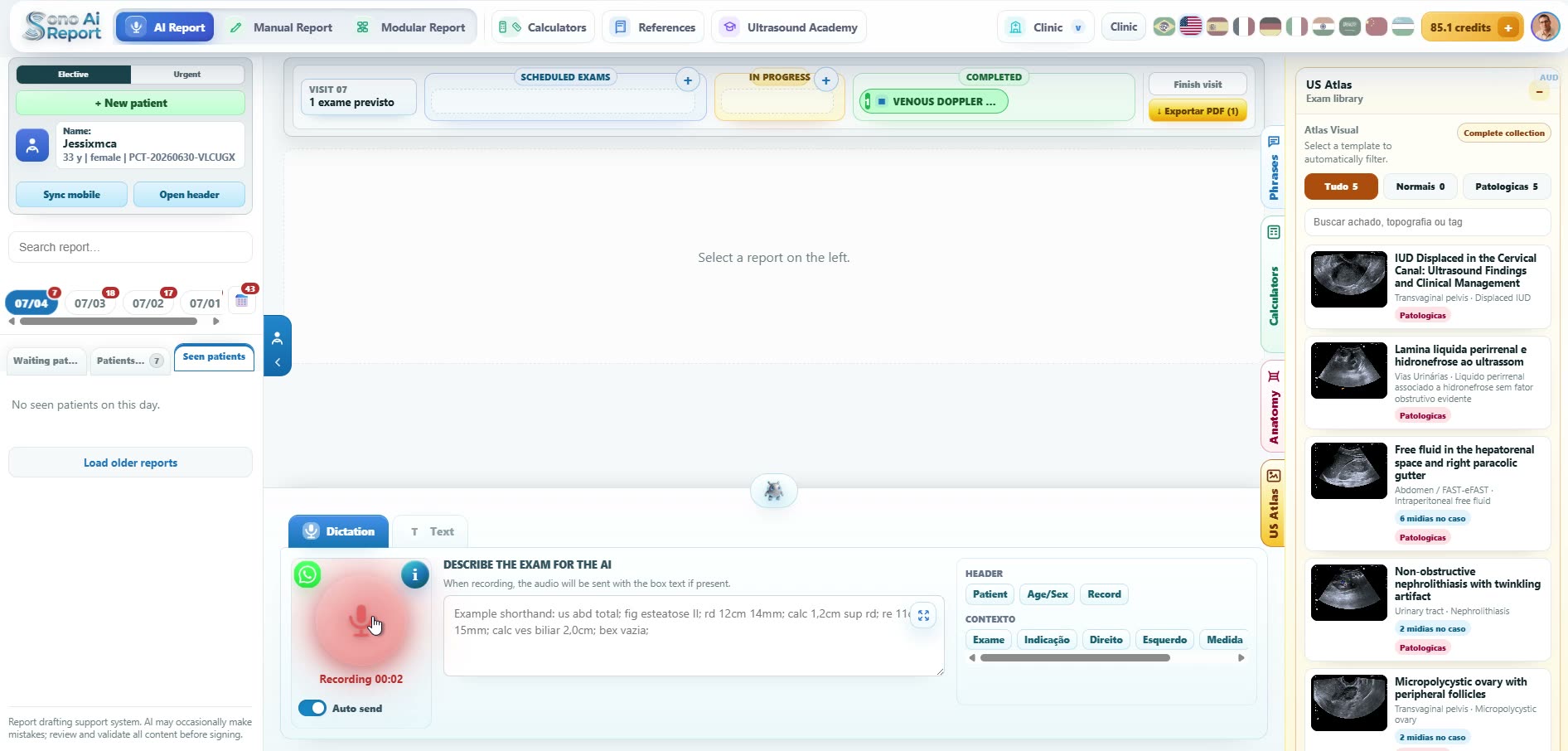
Speak the exam naturally. The engine maps your words onto the platform's standard model, keeps measurements in the body and the opinion in the conclusion, and hands you a finished report to review — the report on the right came from the single dictation on the left.
Dictating · total abdomen
"Total abdomen. Liver with mildly increased echogenicity, grade one steatosis. Simple cyst in segment seven, one point eight centimeters. Gallbladder with a single mobile stone, one point two centimeters, with posterior shadowing. Right kidney with a simple cortical cyst, two point one. Everything else within normal limits."
Ultrasound of the Total Abdomen
Liver: normal dimensions, regular contours and thin edges. Homogeneous parenchyma with mildly increased echogenicity, compatible with grade I hepatic steatosis. Patent hepatic and portal veins of preserved caliber. Highlighting:
Anechoic image with thin walls and posterior acoustic enhancement in segment VII, measuring 1.8 cm — simple cyst.
Bile ducts: intra- and extra-hepatic bile ducts of normal caliber. Common bile duct of preserved caliber.
Gallbladder: normal shape, dimensions and walls, containing a mobile hyperechoic image with posterior acoustic shadowing measuring 1.2 cm, compatible with a calculus.
Pancreas: normal shape, dimensions and contours, homogeneous echotexture, no focal lesions. Main pancreatic duct of normal caliber.
Spleen: normal dimensions, regular contours and homogeneous echotexture, no focal lesions.
Adrenals: adrenal loggias without masses or nodules on the evaluated planes.
Right kidney: topical, normal dimensions, regular contours and preserved corticomedullary differentiation. Highlighting:
Simple anechoic cortical cyst measuring 2.1 cm.
Left kidney: topical, normal dimensions, regular contours and preserved corticomedullary differentiation, without calculi, cysts or pyelocaliceal dilatation.
Abdominal aorta: normal caliber, regular walls, no aneurysmal dilatation.
Inferior vena cava: normal caliber, with preserved physiological respiratory collapse.
Lymph nodes / retroperitoneum: no lymphadenomegaly in the visible retroperitoneal and mesenteric chains.
Free fluid: no free fluid in the abdominal cavity.
Urinary bladder: adequately distended, thin regular walls, anechoic content, no calculi or vegetating lesions.
Bowel loops: without notable changes, no significant wall thickening in the evaluated segments.
Diagnostic impression
- Grade I hepatic steatosis.
- Simple hepatic cyst (segment VII).
- Cholelithiasis.
- Simple cortical cyst in the right kidney.
See it in action
Real screens, real reports.
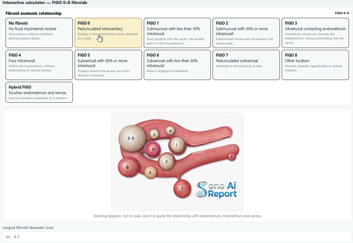
Short, unedited captures from the platform — dictation, integrated classifications and a full modular report.
One environment
Everything a reporting room needs, in one place.
Not a text generator on top of a form — a reporting workflow shaped by daily ultrasound practice.
Voice, AI-text or manual
Three ways to write a report — dictate, assist with AI, or type manually with full control.
Standard exam models
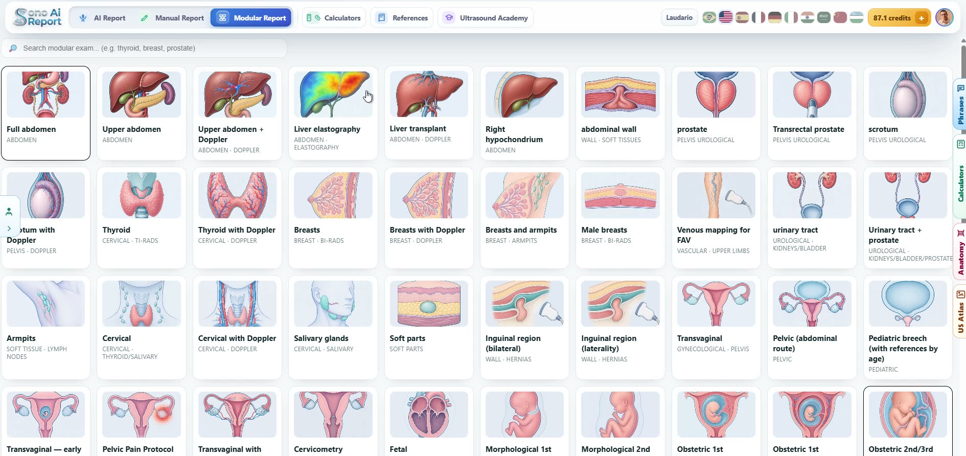
Ready-made modular models per exam — laterality, measurements and conclusions handled consistently. Need your own wording? Build custom models in manual mode.
Calculators built in
TI-RADS, BI-RADS, volumes and indices with results that flow into the report body.
Reference tables on hand
Normal values and measurement ranges one click from the editor — no tab switching.
Branded, verifiable PDFs
Your clinic logo, page-layout presets and a QR verification seal on every report.
Ten languages
Interface and reference content localized — English, Portuguese, Spanish and seven more.
Calculators & references
160+ calculators, scores and reference tables.
Every classification and measurement you reach for, in the same window as the report — results drop straight into the text. No spreadsheets, no second tab.
Modular templates — try them live
Report models built for ultrasound.
Standard modular models per exam: pick a finding and its phrasing loads; add a lesion and the surrounding text adapts so the report never contradicts itself. Click any model below to open it live and build a report yourself — no login. Prefer your own wording? Create custom models in manual reporting mode.
Explore the ecosystem
Two front doors, one source of truth.
Medical hub
For students and reporting physicians: calculators, TI-RADS / BI-RADS classifications, reference values and knowledge to report better — the professional core of the platform.
Open the medical hub →Patient hub
Exam prep and reports explained in plain language — useful when your clinic wants to reassure and inform patients.
Open the patient hub →
Why trust it
Built in a real reading room.
UltrasoundReport.ai is the international home of Sono AI Report, created by Dr. Pedro Pontes — a radiologist who reports ultrasound every day and builds the platform around that routine. Open methodology, published sources, an editorial policy you can read. No black box.
DR. PEDRO PONTES · RADIOLOGIST · CRM 14742 · RQE 9622 · MEMBRO TITULAR DO COLÉGIO BRASILEIRO DE RADIOLOGIA
 LinkedIn
 Ultrasound Academy
Report your next ultrasound with it.
Full platform access during the trial — dictation, calculators, templates and branded delivery.
Start your 7-day free trial →Works in your browser · no card wall to look around